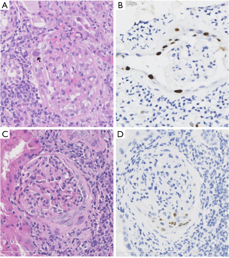
Figure 1.

BK polyomavirus (BKPyV) infection in glomeruli. (A) The BKPyV-infected glomerular parietal epithelial cells presented with typical cytological changes in the peripheral tubular epithelium, with enlarged and ground-glass like nuclei and intra-nuclear inclusions (arrow) [hematoxylin-eosin (HE), ×400]. (B) Immunohistochemistry (IHC) staining showed SV40 large T antigen positivity in the BKPyV-infected glomerular parietal epithelial cells, and the contiguous proximal tubular epithelium (B, IHC, ×400). (C,D) BKPyV infection in both the visceral and parietal epithelium of Bowman’s capsule, with enlarged and hyperchromatic nuclei (C, HE, ×400) and SV40 large T antigen expression (D, IHC, ×400).
