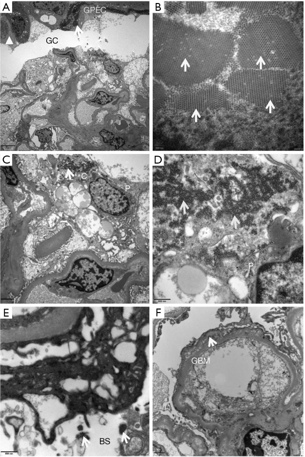
Figure 3.
Ultrastructural changes of BK polyomavirus (BKPyV)-infected glomeruli. The involved glomerular capsule (GC) was thickened and layered (triangle), within which uniform dense structures or membranous cell debris (arrow) was found [A, electron microscopy (EM), ×2,500]. The nuclei of glomerular parietal epithelial cells (GPEC) were enlarged and fusiform, with a large number of virus particles (arrows) (B, EM, ×40,000). Podocytes (P) were occasionally infected by BKPyV. The uniform viral particles (arrows) were found inside the cytoplasm (C, EM, ×6,000), the foot processes (FP) (D, EM, ×20,000), Bowman’s space (BS) (E, EM, ×20,000), and the glomerular basement membrane (GMB) (F, EM, ×10,000).

