Figure 1.
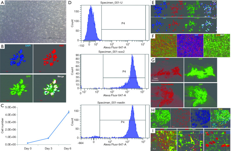
Characterization of induced neural stem cells derived from human fibroblasts. (A) Representative phase contrast images of human induced neural stem cells in proliferation media. (B) Representative fluorescence images of human induced neural stem cells stained for Ki-67 and green fluorescent protein (GFP). (C) Quantification of the cell proliferation rate of human induced neural stem cells maintained under normal conditions for 6 days. (D,E) Representative flow cytometry scatter plots for Nestin- and Sox-2-positive human-induced neural stem cells (D). Representative fluorescence images for Nestin and Sox-2 in undifferentiated neural stem cells (E). (F) Representative fluorescence images of differentiated neural stem cells that were maintained for 14 days in differentiation condition and stained for the neural markers Tuj1, MAP2, and NeuN. (G) Representative images of human induced neural stem cells transplanted into the injured spinal cord. Tissues were stained for human-specific nuclei, Nestin, and green fluorescent protein (GFP) 1 week after transplantation. (H,I) Triple-stained image of human induced neural stem cells transplanted into the injured spinal cord. Tissues were stained for green fluorescent protein (GFP), human-specific Nestin, and MAP2 8 weeks after transplantation (H). Fluorescence image of differentiated neural stem cells transplanted into the injured spinal cord. The GFP-positive neural stem cells were colocalized with MAP2 and synapsin-1. Furthermore, GFP-positive fibers were attached to NeuN-positive neurons (I). Scale bars represent 20 µm (white) and 100 µm (yellow).

