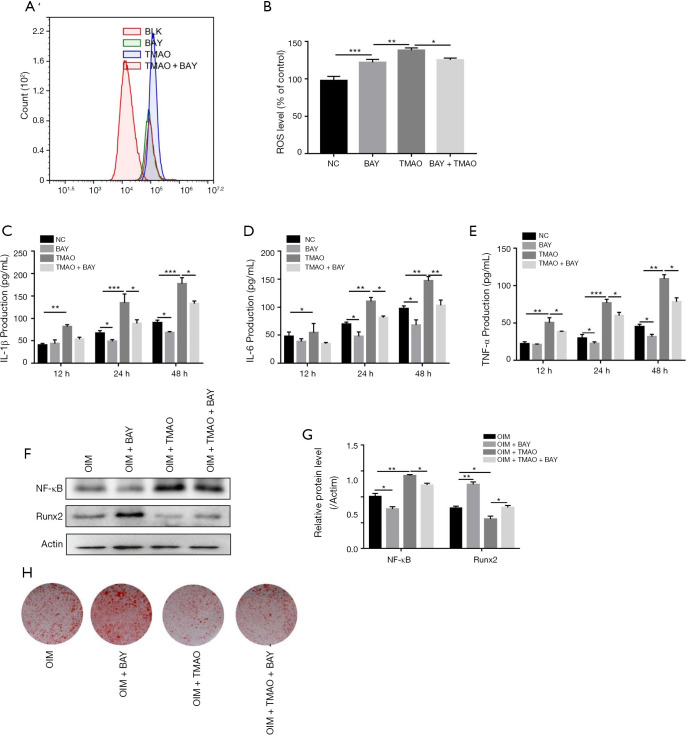
Figure 5.
TMAO regulates BMSC functions via the NF-κB signaling pathway. Cells were pretreated with BAY 11-7082 (10 µM) for 1 h prior to TMAO. (A) Intracellular ROS was detected by flow cytometry; (B) quantification of ROS level was presented as percentage change of mean fluorescence; (C,D,E) the pro-inflammatory cytokine (IL-1β, IL-6 and TNF-α) levels were measured by ELISA assay; (F) WB analysis on NF-κB and Runx2 level in BMSCs cells. Actin was used as an internal control; (G) quantification of NF-κB/Actin and Runx2/actin protein expression; (H) osteogenic differentiation was detected by alizarin red staining. *, P<0.05, **, P<0.01, ***, P<0.001 vs. control group. TMAO, trimethylamine N-oxide; BMSCs, bone marrow mesenchymal stem cells.

