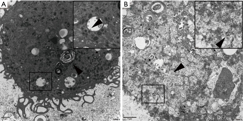
Figure 4.
Electron micrographs of (BALF from a patient infected with SARS-CoV-2. The top right corner shows an enlargement of the selected part of the image. (A) Electron micrographs of BALF from Patient 1showing lamellar bodies in type II alveolar epithelial cells (arrowhead). The virions attached to the smooth vesicles in the cytoplasm (arrowhead). Low magnification (Bar =1 µm, 10,000×), high magnification (Bar =200 nm, 40,000×). (B) Electron micrographs of BALF from Patient 2. The cells were fragmented and the organelles disintegrated; however, the cells, which should be type II alveolar epithelial cells, contained lamellar bodies (arrowhead), and virions were attached to the vesicles (arrowhead). Low magnification (Bar =1 µm, 15,000×), high magnification (Bar =200 nm, 50,000×).

