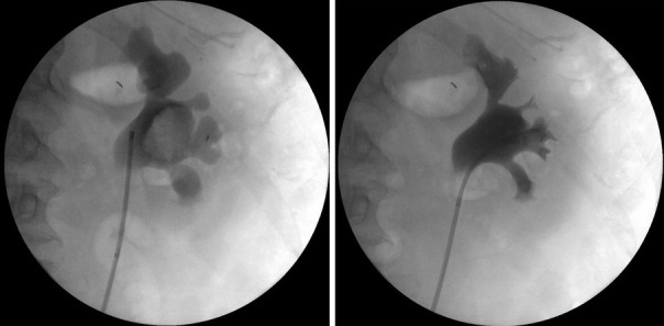
Figure 1.

Example of retrograde uretero-pyelography. Right: retrograde uretero-pyelography of a right upper collecting system. A large filling defect can be visualized in the renal pelvis; left: the same system after endoscopic retrograde treatment. The filling defect is no longer detectable. On the left—tip of catheter inside the UO.
