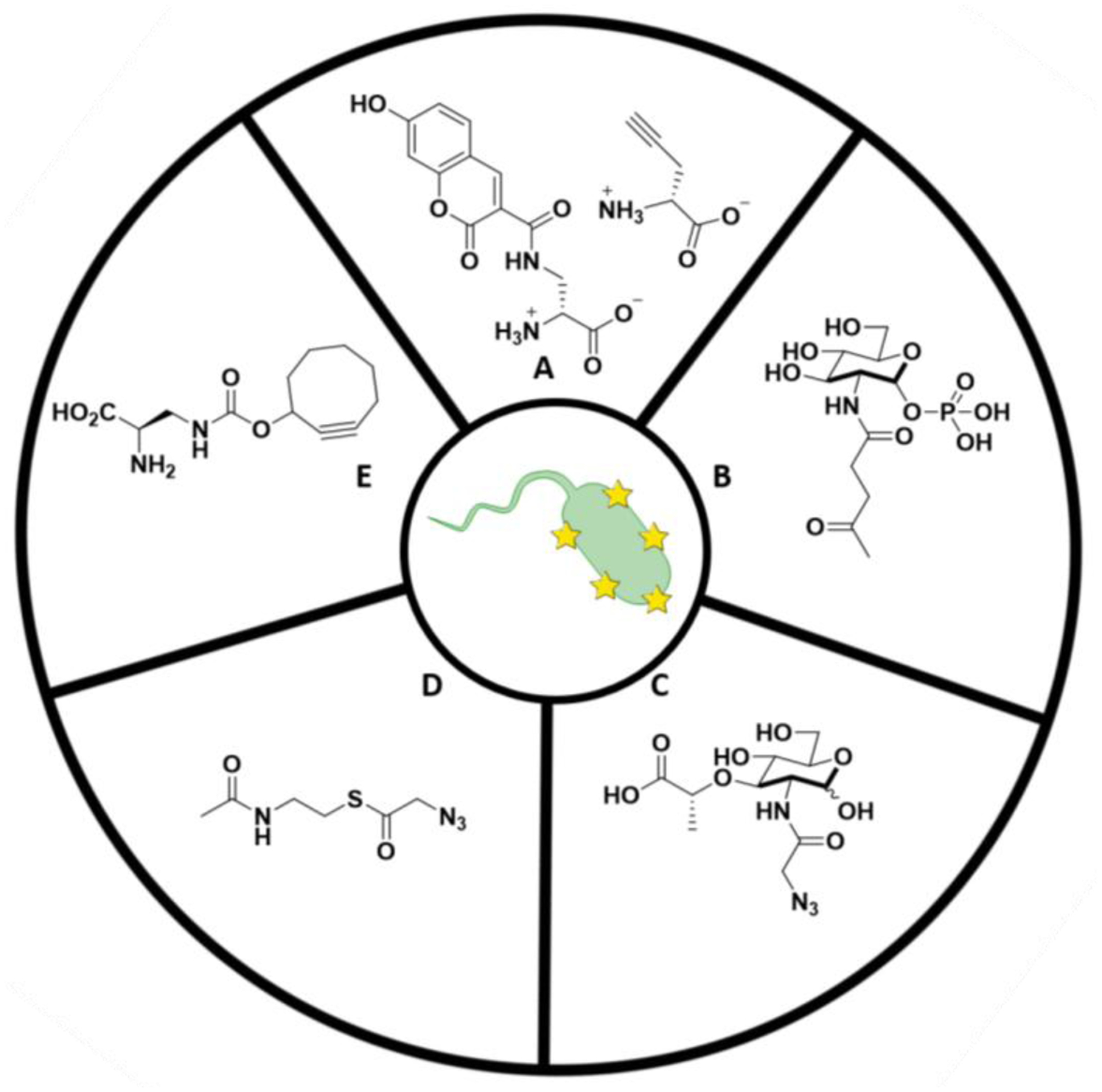
Figure 4: Structure of various bacterial labeling probes.

A) Small fluorophores used to couple to D-amino acids in bacterial PG. B) NAG sugar probe for PG visualization. C) NAM sugar probe for metabolic labeling of PG. D) SNAc derivative example for post-synthetic modification of PG. E) NIR fluorogenic probes for no-wash labeling of PG.
