Figure 3.

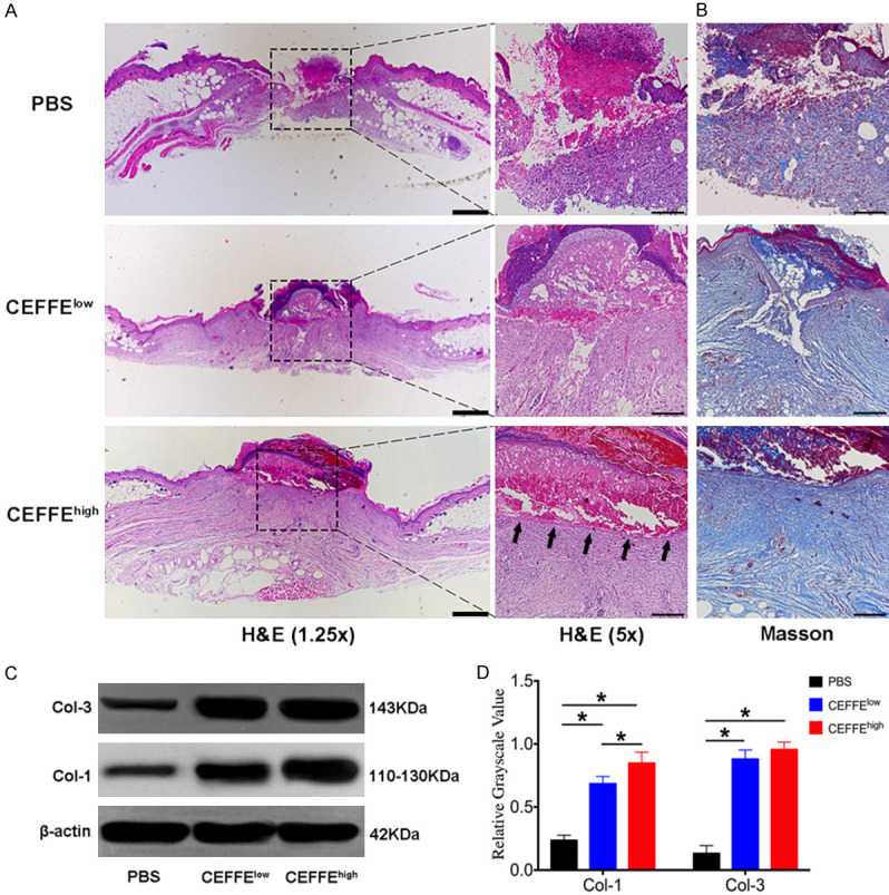
Topical application of CEFFE accelerated cutaneous wound closure, re-epithelialization, and collagen deposition in db/db mice. A. H&E staining for representative wound beds on day 14; re-epithelialization is indicated by arrows. Pictures were taken with a 1.25 × lens (scale bar: 200 μm) and 5 × lens (scale bar: 50 μm). B. Masson’s trichrome staining for representative wound beds after 14 days (collagen deposition is stained blue, scale bar: 50 μm). C. Type I (COL-1) and Type III (COL-3) collagen in the wound beds of each group were measured using the western blot technique. D. Relative quantification of COL-1 and COL-3 in wound beds of each group. Data are represented as mean ± SD; n = 3, *P < 0.05, **P < 0.01.
