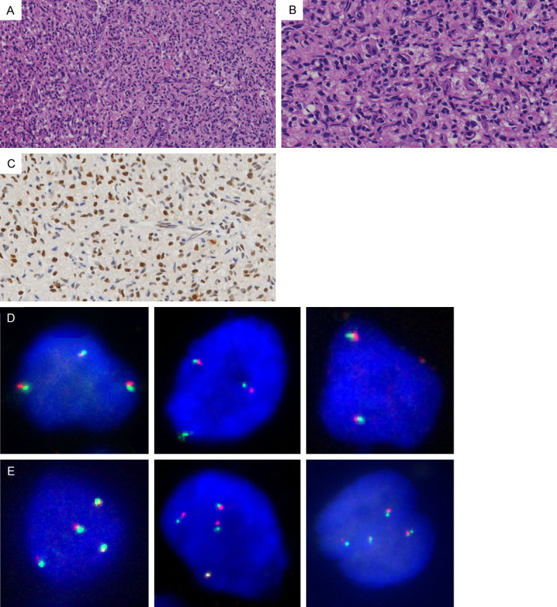
Figure 4.

Histopathological features of HBs harboring polyploid X-chromosome. Representative images from one case of HB. A. Microscopically, the tumor showed high cellularity (H&E; original magnification, 200×). B. The stromal tumor cells contained hyperchromatic nuclei (H&E; original magnification, 400×). C. The stromal tumor cells demonstrated high TFE3 expression (IHC; original magnification, 400×). D. Representative FISH images showing triploid tumor cells (original magnification, 1000×). E. Representative FISH images showing tetraploid tumor cells (original magnification, 1000×).
