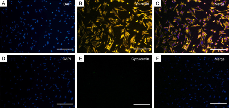
Figure 1.

Typical morphological and immune profiles of uterine ESCs from mice. Immunofluorescence staining in ESCs was positive for vimentin (A-C) and negative for cytokeratin (D-F). Three fields of views were randomly selected under the microscope, and the ratio of positive cells to total cells was counted. The purity of the ESCs population was 96.2% ± 2.5%. Scale bar = 50 μm.
