
Figure 2.
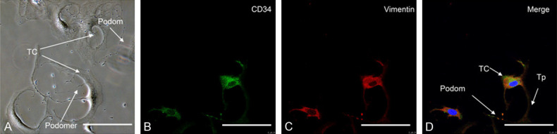
Typical morphological and immune profiles of uterine TCs from mice uterus. A. Phase-contrast limages showed TCs had irregular cell body shapes and characteristic Tps extending from the cell body, with typical alternating podoms (thick segment) and podomers (thin segment). B. CD34 labeled with Alexa Fluor 488 (green). C. Vimentin labeled with (red). D. Merged image showing co-expression of CD34 and vimentin in the whole length of TCs. Representative TCs are shown with, small cell bodies with long Tps, characterized by moniliform outline and alternating podom and podomers. Nuclei were counterstained with DAPI (blue). Scale bar = 50 μm.