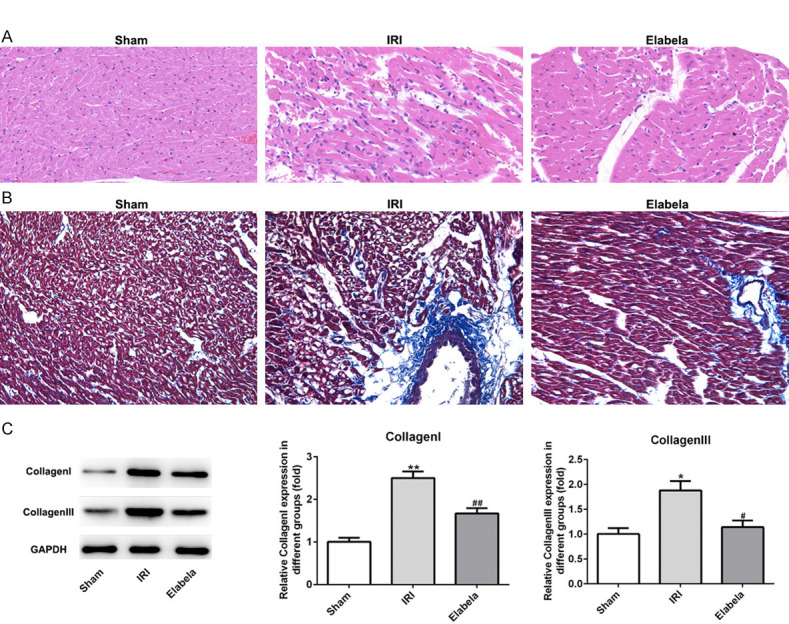
Figure 2.

Elabela decreased cardiac fibrosis after myocardial ischemia/reperfusion injury. A. Heart H&E staining from sham and ischemia reperfusion injury heart tissue. Scale bar: 20 μm. B. Myocardium underwent Masson’s trichrome staining. Red parts indexed cardiomyocytes and blue parts indexed fibrosis. Scale bar: 20 μm. C. Expression levels of type I and type III collagen. IRI, ischemia reperfusion injury. All results were obtained from at least three independent experiments. All numerical data were presented as the mean ± standard deviation. *P < 0.05, **P < 0.01 versus sham group; #P < 0.05, ##P < 0.01 versus IRI group.
