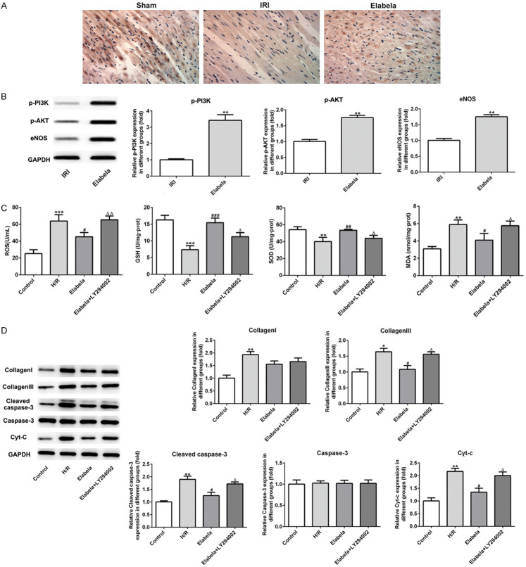
Figure 5.
Elabela modulated the apoptosis and fibrosis by activating PI3K/AKT signaling pathway. A. The expression of phosphorylation-AKT was detected using immunohistochemical staining. B. Expression levels of eNOS, phosphorylation PI3K and AKT were examined using western blot analysis in vivo. **P < 0.01 versus IRI group. H9c2 cells were subjected to high glucose and hypoxia/reperfusion treatment. C. Levels of ROS, GSH, SOD and MDA were detected by commercial kits. D. Levels of type I and type III collagen, cleaved-casepase3, caspase3 and Cyt-c were assessed using western blot analysis in high glucose and hypoxia/reperfusion (H/R)-treated H9c2 cells. All results were obtained from at least three independent experiments. All numerical data were presented as the mean ± standard deviation. *P < 0.05, **P < 0.01, ***P < 0.001 versus control group; #P < 0.05, ##P < 0.01, ###P < 0.001 versus H/R group; ΔP < 0.05 versus Elabela group.

