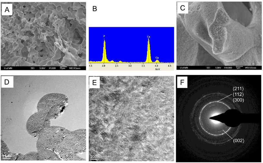
Figure 4.
SEM and TEM analysis of the REDV hydrogel after 14 days of mineralization by the PILP process. (A) SEM image and (B) its corresponding EDS of the mineralized REDV, showing that calcium phosphate minerals were deposited into the REDV hydrogel framework. (C) Fractured surface of the REDV hydrogel after mineralization. (D) TEM image of the mineralized REDV hydrogel showing densely packed and homogeneously distributed minerals in the polymeric matrix. (E and F) TEM image and the corresponding SAED pattern, respectively, revealed that the randomly oritented minerals were needlelike HA nanocrystals.

