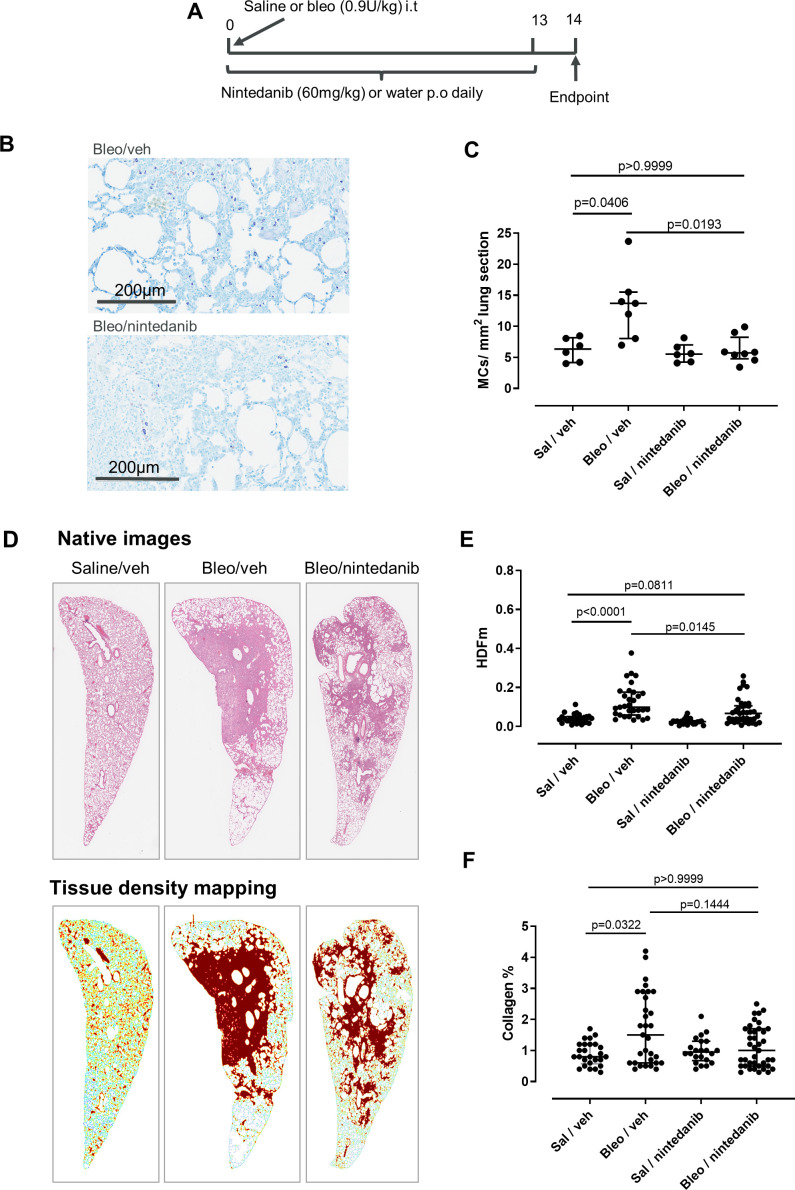
Figure 7.
Nintedanib inhibits lung mast cells (MCs) in the rat bleomycin model. (A) Outline of study design. (B) Rat lungs were stained with Giemsa to visualise the MCs. Representative sections from bleomycin-treated animals treated with nintedanib or vehicle are shown. (C) The number of MCs per area lung was increased with bleomycin and was inhibited by nintedanib. (D) Representative images of annotation of tissue density mapping in which high parenchymal tissue densities specifically located in pulmonary foci have been pseudocoloured in brown. (E) There is a significant increase in the HDFm in the lung parenchyma with bleomycin, and this is inhibited by nintedanib treatment (H&E staining, 5 sections per animal; Sal/vehicle n=6, Bleo/vehicle n=7, Bleo/nintedanib n=9, Sal/nintedanib n=5). (F) There was also a trend towards a decrease of collagen content with nintedanib treatment, although this was not statistically significant (Masson’s trichrome staining, 5 sections per animal) Kruskal-Wallis H=6.571, p=0.0869. Median and IQR are plotted and groups were compared by Kruskal-Wallis with Dunn’s multiple comparisons test.

