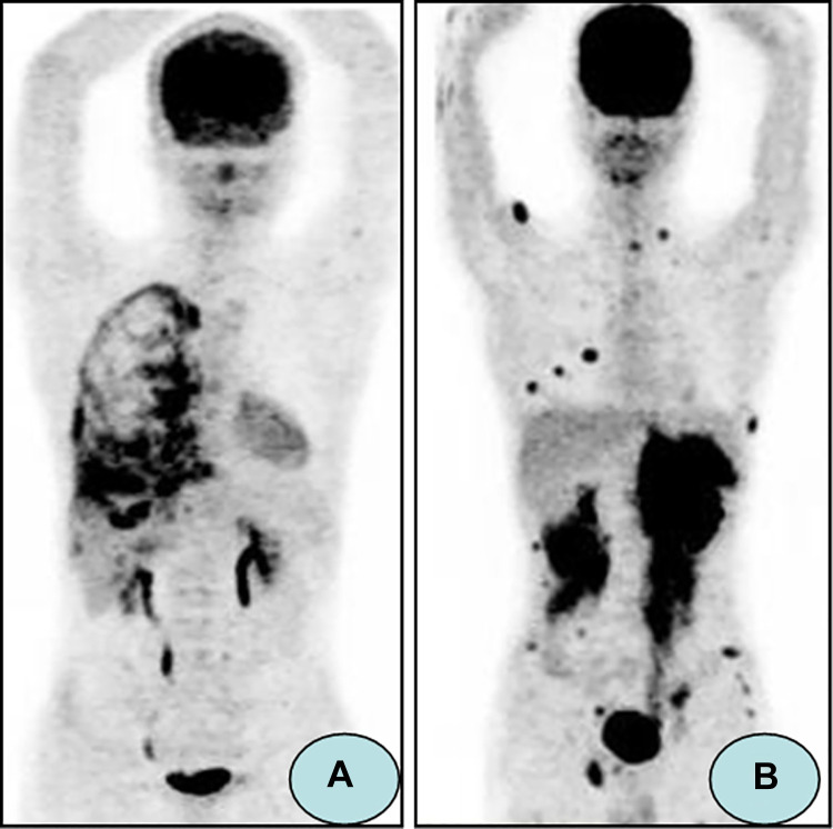
Figure 1.
18FDG-PET/CT images demonstrating extramedullary AL. (A) A 21-year-old male received allo-HSCT for ALL. 18FDG-PET/CT images revealed right pleura with diffuse intense 18F-FDG uptake, which was proved to be extramedullary AL by biopsy. (B) A 20-year-old male received allo-HSCT for ALL. 18FDG-PET/CT images showed bilateral kidneys, muscle, and subcutaneous tissue were involved by extramedullary AL (Reference: Zhou WL, et al.26).

