Figure 2.
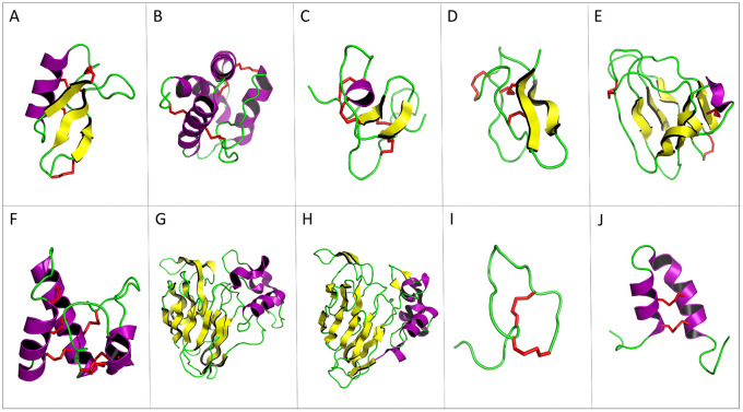
Three-dimensional structure of plant antimicrobial peptide representatives. Yellow arrows correspond to β-sheets; α-helices are represented in purple and disulfide bonds in red. (A) Defensin NaD1 of Nicotiana alata (PDB ID: 1MR4). (B) Lipid transfer protein TaLTP1.1 isolated from wheat (PDB ID: 1GH1). (C) Hevein of Hevea brasiliensis (PDB ID: 1HEV). (D) Knottin Ep-AMP1 of Echinopsis pachanoi (PDB ID: 2MFS). (E) MiAMP1 peptide of Macadamia integrifolia (PDB ID: 1C01). (F) Snakin-1 of Solanum tuberosum (PDB ID: 5E5Q). (G) Thaumatin from Thaumatococcus daniellii (1RQW). (H) Zeamatin of Zea mays (1DU5, chain A). (I). Theoretical 3-dimensional model of Impatiens balsamina IbAMP1. (J) Helical hairpin structure of the novel antimicrobial peptide EcAMP1 from seeds of barnyard grass (Echinochloa crus-galli) (PDB ID: 2L2R). PDB indicates protein data bank.

