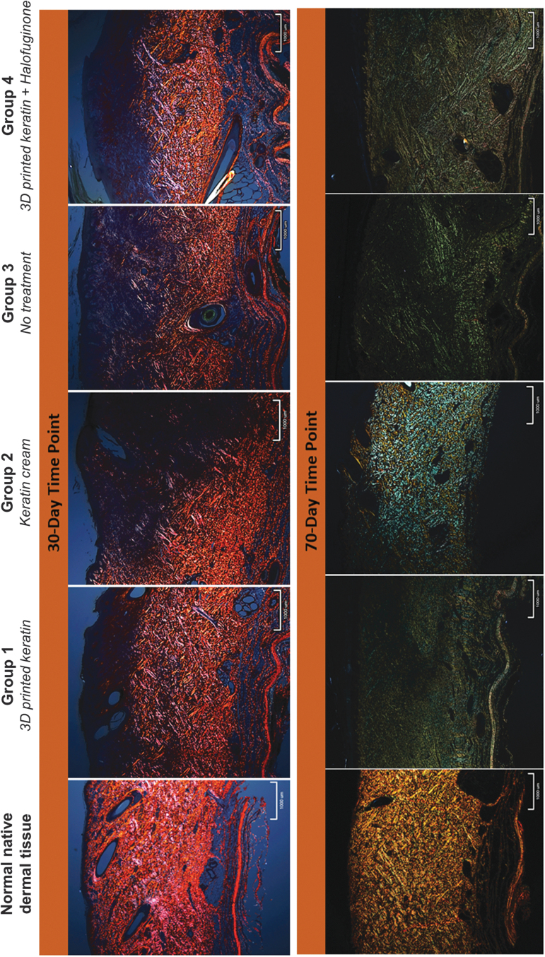
FIG. 5.
Representative (low-power, × 20) sections of Picrosirius Red-stained samples of all treatment groups imaged under polarized light. Collagen content and order is characterized by the intensity and pattern of polarized light-induced birefringence of collagen fibers. Intense red/orange/yellowish birefringence indicates mature and organized collagen fibers (control, normal tissue, far left). Green birefringence has generally been correlated to immature, fine, and less organized collagen, although color change can be correlated to rotations of the samples and light incidence.51 In this study, color was not considered to assess quality of the collagen but merely to identify it and its general orientation trends. At 30 days postprocedure, the heterogeneity of coloration is indication of collagen orienting in different directions (black, purple, orange, and red are indicative of different orientation); collagen is only present but disorganized in the lower reticular dermis. At 70 days, images show improved patterns or heterogeneity, with collagen present throughout the dermal layers although still disorganized with different orientations colored blue/green and yellow. Scale shown: 1 mm.

