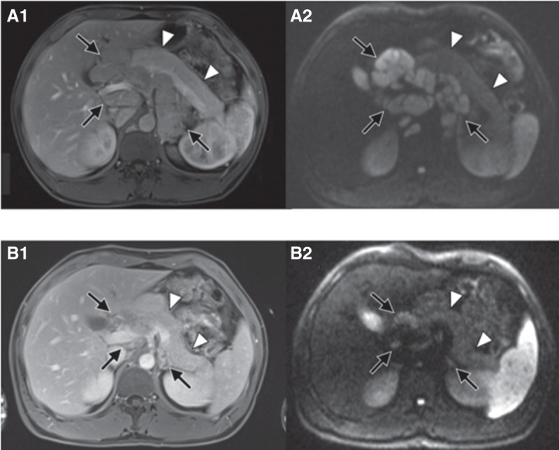
Figure 3.

Almost complete remission at 3-mo follow-up after initiation of trametinib/dabrafenib. Baseline imaging: axial early venous phase MR image (A1) and axial diffusion weighted MR image with b-value of 800 sec/mm2 (A2) show extensive lymphadenopathy in the retroperitoneum and liver hilum (black arrows). There is no lesion in the pancreas (white arrowheads). Follow-up imaging: Axial early venous phase MR image (B1) and axial diffusion weighted MR image with b-value of 800 sec/mm2 (B2) show marked reduction of lymphadenopathy (black arrows). Still there is no lesion in the pancreas (white arrowheads).
