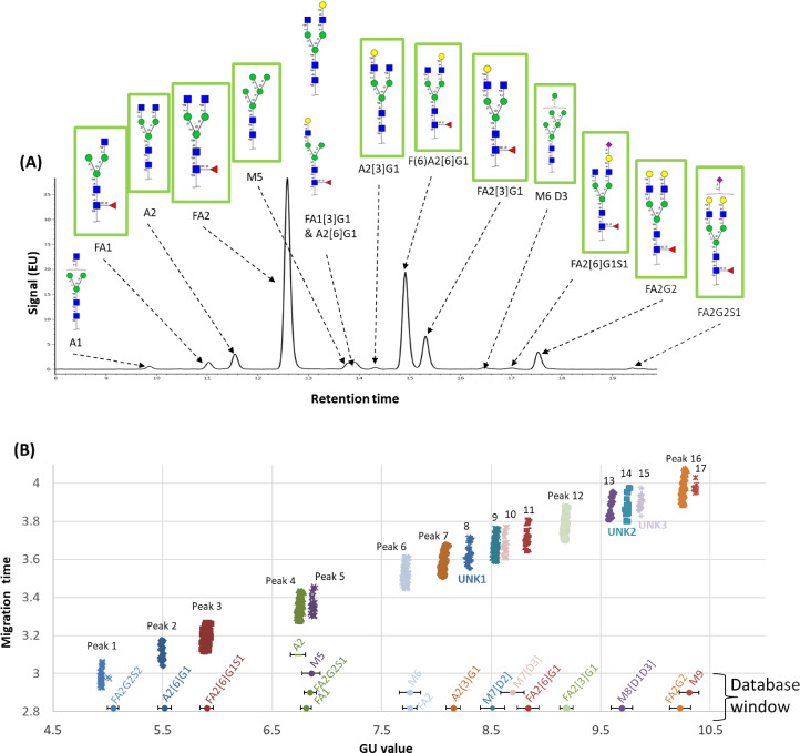
Figure 5.
Glycans identified in anti-HER-2 samples using UPLC-MS and CE. (A) the UPLC chromatogram confirmed the 14 glycans using GU and mass. Green boxed glycans were also identified in CE. (B) Glucose units vs. migration time for all 391 CE electropherograms. Database matched glycans are shown in Oxford linear notation [19]. The CE APTS database hits are marked with a circle and a corresponding error bar showing the GU tolerance. All glycans with core fucose were α-1→6 linkage, galactose were β-1→4 linkage and all sialic acid linkages were α-2→3 linkage. All glycans are drawn in SNFG notation [20].

