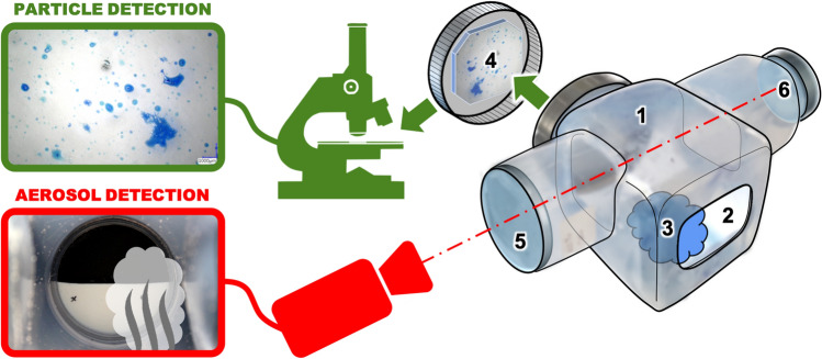
Fig. 1.
Representation of the sample chamber (1) with an opening (2) for processing the sample material (3). Ejected particles are collected on the slide (4) on the rear wall for later microscopic analysis. In the upper part of the sample chamber the aerosol formation is video-documented through an observation tube (5). Here the haze of the view of a target object (6) is evaluated

