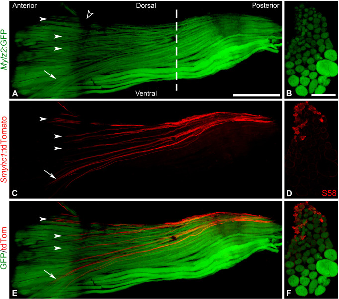
Figure 1.
The myofiber organization of the zebrafish MR muscle. Longitudinal (A, C, E) and cross-sectional (B, D, F) view of adult whole-mount MR expressing mylz2:GFP (fast, A, B), smyhc1:tdTomato (slow, C, D), and merged (E, F). Open arrowhead indicates a shade created by the sclera, white arrowheads indicate slow myofibers (smyhc1:tdTom positive) spreading across the muscle, and the white arrow indicates a subgroup of myofibers reaching diagonally across the muscle. The dashed line indicates approximate level where cross sections of the MR shown were cut in B, D, F. Scale bar in A applies also to C and E: 500 µm; scale bar in B applies also to D and F: 50 µm.

