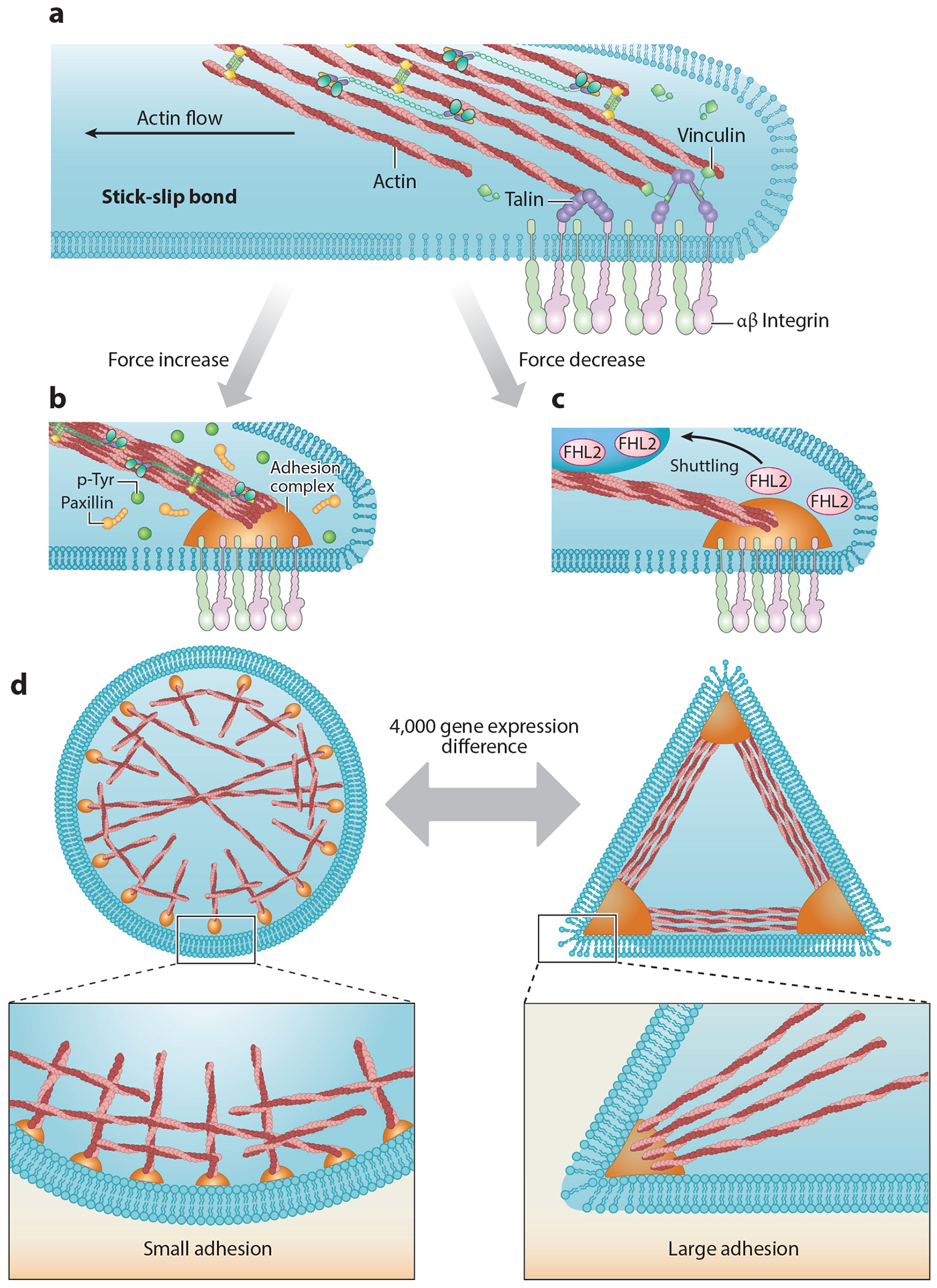
Figure 3.

(a) Established adhesions experience drag forces from actin flowing to the cell center, resulting in a stick-slip behavior at the actin-adhesion interface (68). At later time points, when the adhesions mature, actin drag forces lead to repeated talin stretch-relaxation events that involve the binding of several vinculin molecules to a single talin (7). (b) Increasing forces cause increases in local tyrosine phosphorylation levels and reinforce adhesion formation. (c) Decreasing force leads to FHL2 shuttling to the nucleus (63). (d) Within 4 h of plating, cells on circular patterns show different expression levels of more than 4,000 genes compared with cells on triangle patterns (19). Abbreviations: FHL2, four and a half LIM domains protein 2; p-Tyr, phosphotyrosine.
