
Figure 1.
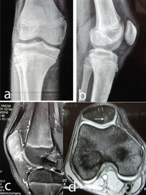
(a-d) Anteroposterior and lateral radiographs of the knee in a young male with anterior knee pain. No gross abnormality was detectable. (c) T2-weighted sagittal magnetic resonance imaging (MRI) section showing patellar edema. (d) T2-weighted axial MRI section showing a hypointense punctate lesion surrounded by a small hyperintense zone near the medial border of patella, close to its articular surface (arrow).