Figure 2.

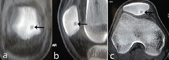
(a and b) Computed tomography scan of the patient (a) coronal reformat, (b) sagittal reformat, and (c) axial cut showing central nidus surrounded by minimal sclerosis.

(a and b) Computed tomography scan of the patient (a) coronal reformat, (b) sagittal reformat, and (c) axial cut showing central nidus surrounded by minimal sclerosis.