Figure 3.

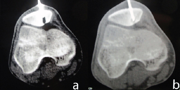
(a and b) Percutaneous computed tomography-guided radiofrequency ablation (a) insertion of guide wire through the dorsal approach, (b) the guide wire is in position and would be replaced by the radiofrequency probe.

(a and b) Percutaneous computed tomography-guided radiofrequency ablation (a) insertion of guide wire through the dorsal approach, (b) the guide wire is in position and would be replaced by the radiofrequency probe.