Figure 5.

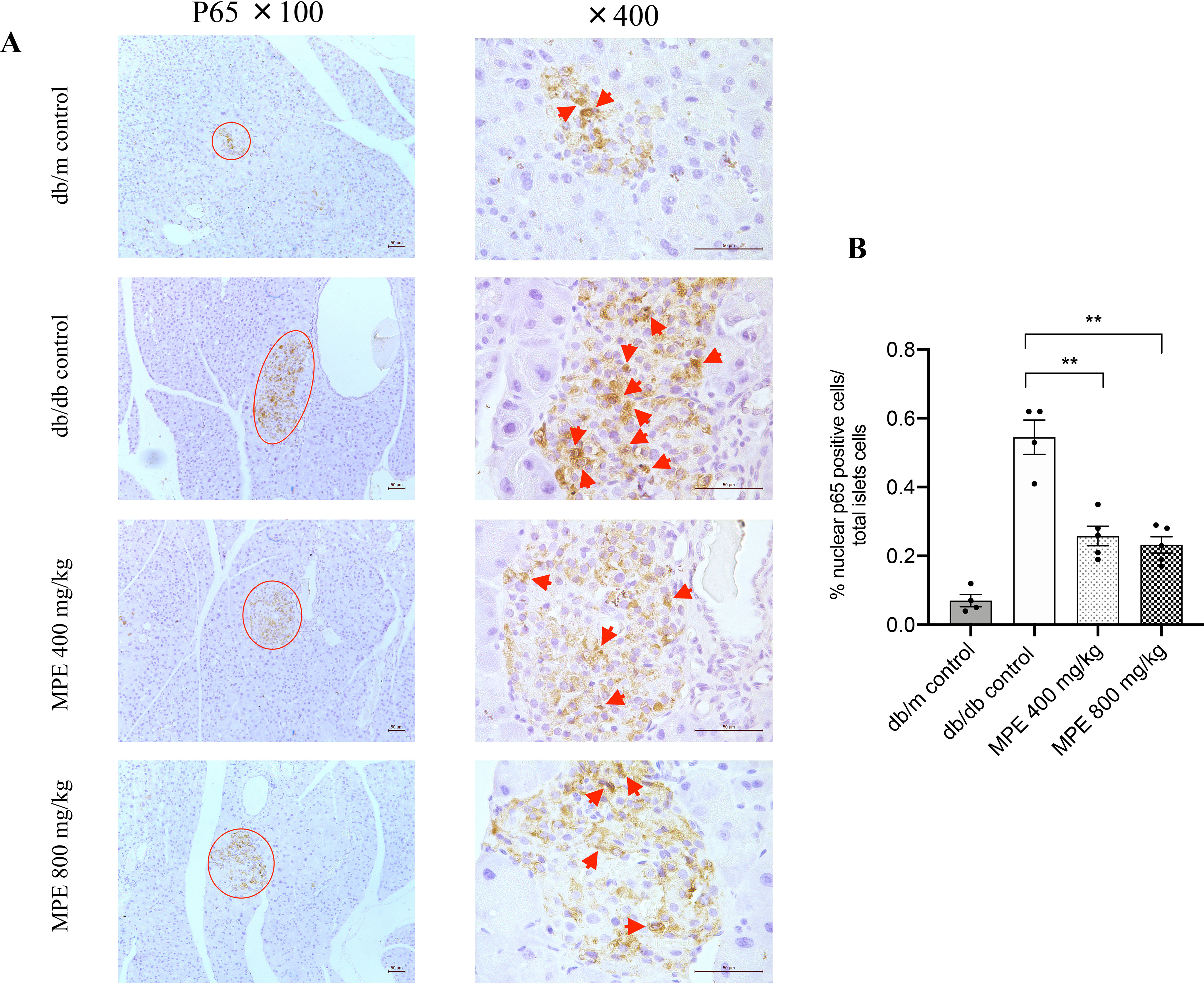
Impact of MPE on NF-κB p65 nuclear staining and IL-1β expression. A, immunohistochemistry staining of p65 in the nucleus of pancreas of db/db mice (magnification: ×100 and ×400; both the short and long black scale bars in the top panel in the bottom right corner represent 50 μm, respectively). Red arrow, positive signal in the islet. B, relative quantification of p65-stained positive signal in the pancreas of db/db mice. C, real-time PCR analysis of relative il1b mRNA expression levels in MIN6 cells with treatment of MPE or control in high-glucose (25 mmol/liter) or low-glucose (5.5 mmol/liter) medium (LG control). D, IL-1β protein levels in supernatant after treatment of MIN6 cells with IL-1β or IL-1β + TNFα + IFNγ. E, real-time PCR analysis of relative il1b mRNA expression levels in primary islets from HFD-induced obese mice with treatment of MPE or HG control. F, IL-1β protein levels in supernatant after treatment of primary islets of HFD-induced obese mice with IL-1β or IL-1β +TNFα + IFNγ. G, GSIS was performed to evaluate the primary islet function from C57 mice with normal chow feeding after treatment with IL-1β or IL-1β +TNFα + IFNγ in LG control and MPE groups. H, real-time PCR analysis of relative il1b mRNA expression levels were measured after treatment with IL-1β or IL-1β + TNFα + IFNγ in primary islets of C57 mice in LG control and MPE groups. Data are presented as mean ± S.E. (error bars) with individual data points in histograms. *, p < 0.05; **, p < 0.01.
