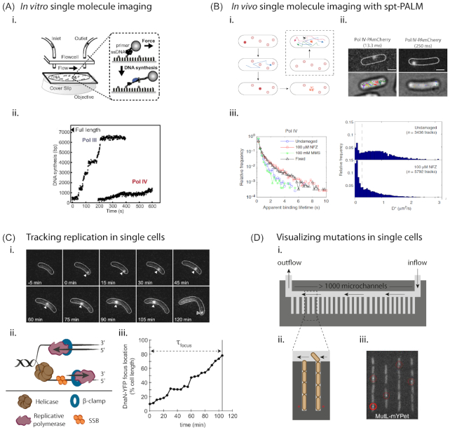
Figure 1.
Visualizing repair across scales. (A)In vitro single-molecule imaging of TLS polymerase activity. (i) Schematic showing the setup used by Kath et al. (2014), for visualizing polymerase switching. (ii) Representative trajectories showing Pol IV or Pol III synthesis over time on individual DNA molecules (Kath et al. 2014). Images reprinted with permission from PNAS.(B)In vivo single-molecule imaging of TLS activity. (i) Cartoon representation of the method of single-particle tracking with photoactivable localization microscopy (spt-PALM) (adapted from Stracy et al. 2014). Photoconvertible fluorophores are used and only one molecule is activated per cell at any point in time. These molecules are then imaged over time to capture single-molecule trajectories. Populations of freely diffusing, slow diffusing and bound molecules are identified and their characteristics studied. (ii) and (iii) Application of spt-PALM to study Pol IV activity in E. coli reveals association of Pol IV with DNA under damage-induced conditions. Images reprinted with permission from Thrall et al. (2017) and shared under Creative Commons public license (creativecommons.org/policies). (C) Tracking replication in single cells (from Aakre et al. 2013). (i) In this example, the β-clamp (DnaN) in Caulobacter crescentus is fluorescently tagged with YFP. Replication initiates at one cell pole and is tracked over time until completion at the opposite cell pole, where DnaN dissociates from DNA and its localization is lost. (ii) Schematic representation of the replication fork. (iii) DnaN position over time for cell in (i) is shown. τfocus represents time from focus formation to loss (Aakre et al. 2013). Image reproduced with permission from Elsevier. (D) Visualizing mutations in single cells. (i) and (ii) Microfluidics devices such as the ‘mother-machine’ allow the visualization of >105 cells in a single experiment. This can be combined with fluorescence imaging to follow activity and regulation of repair pathways, such as TLS, in single cells. (iii) This approach has been used to track mutations in real-time by following MutL-YFP foci that denote the position of DNA mismatches (Uphoff 2018). Image reproduced with permission from PNAS.

