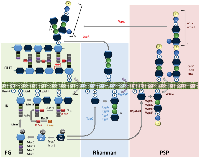
Figure 2.
Schematic representation of the main proposed steps for the biosynthesis of peptidoglycan (PG, green background) and cell wall polysaccharides (CWPS), including rhamnan (blue background) and polysaccharide pellicle (PSP, pink background) in L. lactis. Hexagons represent MurNAc (light blue) and GlcNAc (dark blue). Circles represent sugars: rhamnose (green), glucose (blue), galactofuranose (yellow). Rectangles represent amino acids: L-Ala (black), D-Glu (green), L-Lys (purple), D-Ala (grey), D-Asp (red). P, phosphate. The biosynthesis of the three cell wall glycopolymers starts in the cytoplasm (IN). For PG, a soluble precursor, UDP-MurNAc-pentapeptide is first synthesized then transferred onto the lipid carrier undecaprenyl-phosphate (Und-P) by MraY (forming lipid I) and further assembled by MurG to form the lipid II precursor. The rhamnan chain and the PSP subunit are both assembled on the lipid carrier, Und-P. In our model, rhamnan synthesis is initiated by the transfer of GlcNAc-P onto Und-P by TagO, whereas PSP subunit synthesis is initiated by the transfer of GlcNAc onto Und-P. The three lipid-bound precursors are translocated to the outer side of the cytoplasmic membrane. MurJ is the flippase involved in the translocation of lipid II and WpsG is the presumed flippase involved in the export of lipid-linked PSP subunit. RgpC/D is an ABC-transporter involved in the transport of lipid-linked rhamnan chains. At the outer side of the membrane (OUT), PG subunits are polymerized by PBPs and possibly by SEDS (shape, elongation, division and sporulation) proteins (not shown). LcpA is proposed as the main transferase involved in anchoring rhamnan onto PG and WpsJ is a membrane glycosyltranferase with a GT-C fold proposed to be involved in attaching PSP onto rhamnan. The nature of the bond between PSP and rhamnan chains is unknown. The three proteins CsdC, CflA and CsdD are involved in the addition of side chain Glc onto PSP subunits, most probably at the outer face of the membrane. See text for further details. This figure is adapted from Sadovskaya et al. 2017 and Theodorou et al. 2019.

