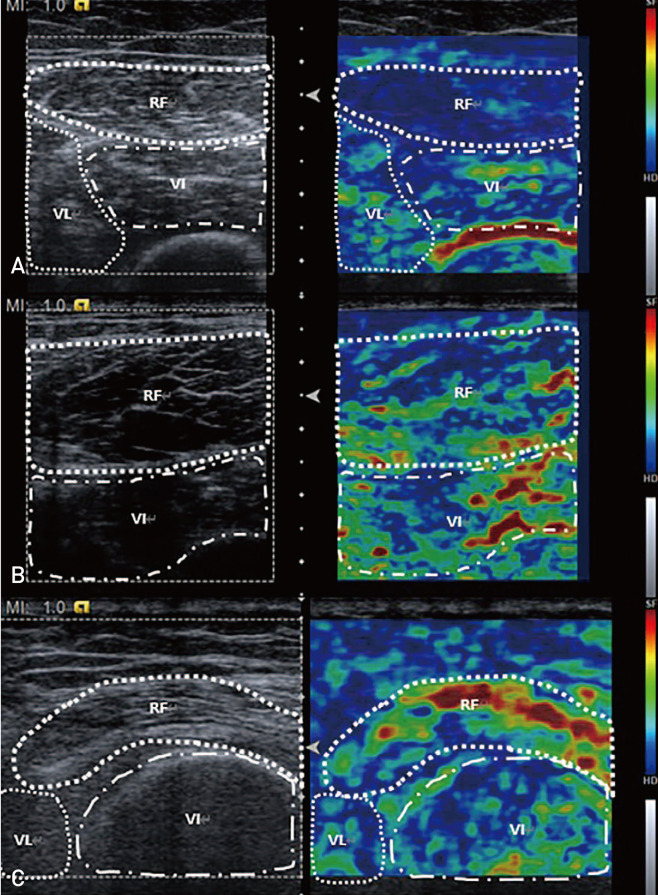
Fig. 1. Conventional ultrasonographic (US) images of the rectus femoris muscle and sonoelastographic images using frozen US images. (A) The blue region is dominant and thus classified as grade 0. (B) When The blue region is relatively dominant (i.e., involved more than half of the muscle), it is classified as grade 1. (C) When the area occupied by the blue region is less than half, it is classified as grade 2.
RF: rectus femoris, VL: vastus lateralis, VI: vastus intermedius.

