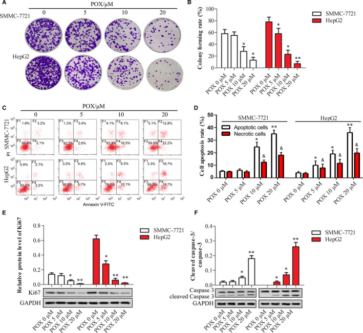
FIGURE 2.

Puerarin 6″‐O‐xyloside inhibited proliferation and promoted apoptosis in SMMC‐7721 and HepG2 cells. Both cell lines were treated with 0, 5, 10, and 20 μmol/L of Puerarin 6″‐O‐xyloside for 24 h. The proliferation was shown with crystal violet staining (A) and area of the staining was measured by ImageJ (B). Flow cytometry was applied to show apoptotic cells (C) and the number of apoptotic cells was counted (D). Western blotting was used to show relative protein expression of Ki67 (E) and the ratio of cleaved caspase‐3 to caspase‐3 (F) in both cell lines. *P < .05 and **P < .01 compared with no Puerarin 6″‐O‐xyloside treatment by t test of at least three replicates. POX: Puerarin 6″‐O‐xyloside
