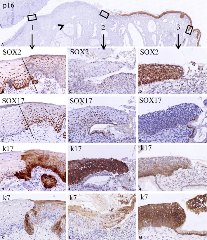
FIGURE 4.

Switches in the expression of SOX2 and SOX17 in the transition of normal epithelium to high‐grade squamous intraepithelial lesions (HSIL). Low magnification of a section stained for p16 showing negative normal epithelia and a positive HSIL lesion (A). The boxes 1‐3 in this figure indicate epithelial transitions within the normal epithelium (box 1: a transition between normal and metaplastic squamous epithelium, possibly the original squamocolumnar junction), a new squamocolumnar junction (box 2), and the transition between HSIL and glandular epithelium (box 3). The fact that glands are seen beneath the squamous epithelium between boxes 1 and 2 (arrowhead) indicates that this region can be regarded as the transformation zone. The higher magnifications of these areas (B‐M) illustrate the mutually exclusive expression of SOX2 and SOX17 in these transitions. This is again evident in the keratin 17‐ and keratin 7‐negative normal ectocervical squamous epithelium in the p16‐negative area (left epithelial area in B, E, H, K). The keratin 17‐ and keratin 7‐positive metaplastic epithelium (right epithelial area in B, E, H, K) shows an extensive SOX17 positivity in the basal and intermediate compartment, and in this case SOX2 positivity in the superficial layers. In the strongly keratin 17‐positive squamous epithelium close to the new SqCJ only SOX17 is expressed, while SOX2 expression is absent (C, F, I, L). This SOX expression pattern is reversed in HSIL as identified by expression of keratins 7 and 17, where SOX2 expression is high and SOX17 expression is absent (D, G, J, M; p16 positive area)
