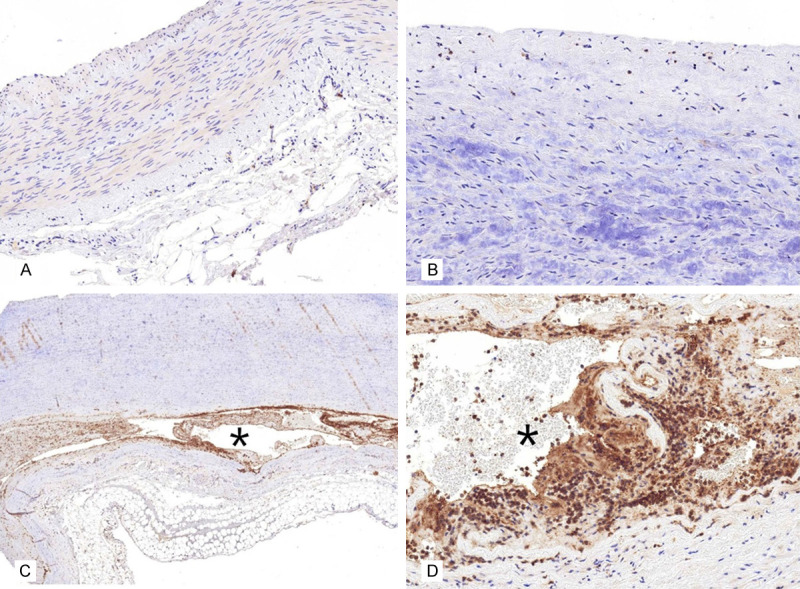
Figure 5.

Caspase 3 showed negative immunohistochemical staining result in the normal (A. ×200) and atherosclerotic (B. ×400) aorta. By contrast, caspase 3 showed strongly positive expression levels (C. ×100) in the region close to the dissected lesion (asterisk) and in the cytoplasm of smooth muscle cells (D. ×400) near the dissected lesion (asterisk).
