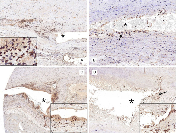
Figure 6.
Immunohistochemistry for p-Smad3/4, Snail/Slug, and TGF-β1. A. The expression levels of p-Smad3/4 are weakly positive near the dissected lesion (asterisk). The inset shows the inflammatory and smooth muscle cells (SMCs) stained with p-Smad3/4 near the dissected lesion (×200). B. The p-Smad3/4 expression levels are slightly stronger in the fibrotic area (arrow) near the dissected lesion than in the control atherosclerotic aorta (×400). C. The expression levels of Snail/Slug are weakly positive in the nuclei of the inflammatory cells and SMCs (inset, ×100) near the dissected lesion (asterisk). D. The TGF-β1 expression levels are slightly stronger (arrow and inset, ×100) in the region near the dissected lesion (asterisk) than in other regions of the aortic media.

