Fig. 3.
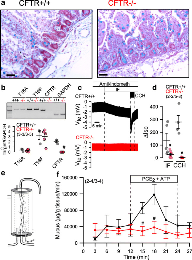
Lack of intestinal fluid and mucus transport in CFTR−/− piglets. a Histology and Alcian blue staining of intestinal epithelium from CFTR+/+ and CFTR−/− piglets. Bars = 100 μM. Representative stainings from n = 2 CFTR+/+ and CFTR−/−, respectively, with 22–23 images being analyzed. b Expression of TMEM16A, TMEM16F, and CFTR in intestinal epithelial cells from CFTR+/+ and CFTR−/− piglets as detected by RT-PCR. c Original Ussing chamber recordings of transepithelial voltages (open circuit) from freshly isolated jejunal epithelium obtained from CFTR+/+ (upper recoding; black) and CFTR−/− (lower recording; red) piglets. d Comparison of calculated short circuit currents activated by basolateral IBMX (100 μM) and forskolin (2 μM) (IF) or the muscarinic agonist carbachol (CCH, 100 μM). e Scheme for the perfusion of isolated CFTR+/+ and CFTR−/− jejunum ex vivo (left) and f time course for mucus release induced by luminal perfusion with PGE2 (1 μM) and ATP (100 μM). Mean ± SEM (number of animals/number of experiments or reactions). #Significant difference when compared with CFTR+/+ (p < 0.05, ANOVA)

