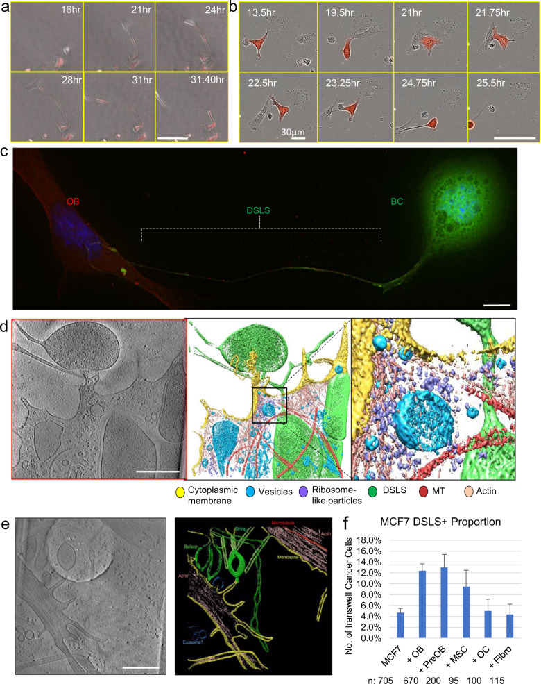
Fig. 2. Luminal breast cancer cells form dendritic spine like protrusions that tether them to mobile osteoblasts.
a Time-lapse on Cytation of mobile osteoblasts contacting and drawing out cellular protrusion from MCF-7 (red). Scale bar is 100 µm. b Time-lapse on Incucyte S3 of osteoblast (unlabeled) drawing out cellular tether protrusion that leads to MCF-7 (RFP-labeled,) migration. Scale bar is 100 µm. c Deconvolution imaging of MCF-7 cell (green) with dendritic spine-like structures (DSLS) protrusions attached to OBs (red). Scale bar is 10 µm. d Tomogram of the terminus of DSLS (green) touching OB cell membrane (yellow). Detail of actin and ribosome concentration where cells make contact. Scale bar is 500 nm. e Tomogram of multiple DSLS structures and actin within. Scale bar is 500 nm. f The relative abundance of MCF-7 DSLS structures in co-culture with various bone cells, normalized to abundance in monoculture. OB: osteoblasts represented by hFOB 1.19 cells. PreOB: Pre-osteoblasts represented by MC3T3-E1 cells. OC: osteoclasts represented by RAW 264.7 cells. MSC: immortalized Mesenchymal Stem cells Fibro: fibroblasts represented by 293T cells. Error bars indicate standard deviation calculated based on binomial distribution (see “Methods”).

