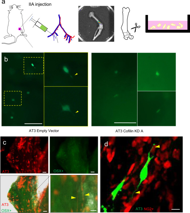
Fig. 5. Evidence of DSLS in breast cancer bone metastasis.
Labeled AT3 cells were injected via intra-iliac artery (IIA) injection into C57B/6 mice. Mice were immediately sacrificed, hind limb bones harvested, and cut into small (1–2 mm diameter) pieces. Bone fragments are cultured over the next 3 days and imaged each day. a AT3 cells with both empty vector and cofilin knockdown (KD) shRNA transfection in the bone 24 h after intra-iliac artery injection, sacrifice, and bone culture. Cofilin knockdown decreased the proportion of DTCs with visible DSLS from 50% to 16.7% (n = 9 mice). Yellow arrows indicate potential DSLS. Scale bars are 150 µm. b Spontaneous bone metastasis generated by orthotopic mammary tumors of RFP-tagged AT3. Hosts were OSX-GFP mouse mice. Red, green and merged channels are shown separately. Yellow arrows indicate potential DSLS in the vicinity of OSX+ cells. Scale bars are 50 (upper left), 50 (upper right), 50 (lower left), and 20 µm (lower right). c NG2-RFP mouse bone fragment containing GFP-tagged AT3 cells delivered by intra-iliac artery injection. Yellow arrows indicate DSLS from RFP-tagged AT3 cells terminating in contact with NG2+ mesenchymal stem cells. Scale bar is 15 µm.

