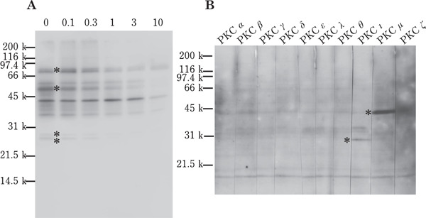
Fig. 3.

Western blot analysis of sperm lysates. (A) Ejaculated sperm were incubated with BisII (0, 0.1, 0.3, 1, 3 or 10 µM) for 30 min and the sperm lysates were separated by SDS-PAGE followed by Western blot analysis using anti-phospho-PKC substrate antibody. The intensity of the bands indicated by asterisk in lane 0 was decreased by the addition of 1 µM BisII. (B) The lysates of fresh ejaculates were separated by SDS-PAGE and detected with anti-PKC antibodies. The immunoreactive band in lane PKCι and PKCµ was indicated by asterisk. A representative image of repeated experiments is shown.
