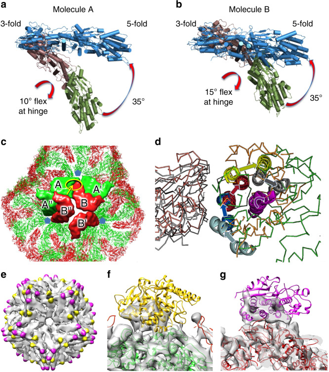
Fig. 3. Rearrangement between SLP and virion-like particles.
a, b Side views of the conformational changes within the λ1 subunits between virion-like particle (blue) and the SLP where the two portions are coloured raspberry and forest, the latter portion being closest to the fivefold. c Interface areas between λ1 molecules in the SLP. Highlighted in yellow is the area shown in (d) with the arrow indicating the viewpoint. d Close up view of the ratchet of the AB interface. The A molecules are on the left. SLP molecules A and B coloured grey and orange, respectively. Molecules A and B for the virion-like particle are coloured pink and green, respectively. To show the ratchet more clearly the A molecules of each AB pair have been superposed. For the B molecules ratchetting helices and the pivot helix are coloured equivalently for the SLP and the virus (the pivot helix is shown in purple). Arrows show the rotation between the SLP and virus. e SLP density (PEET map) unaccounted for by λ1. Within the icosahedral asymmetric unit there are two distinct sets of density, one close to the twofold is coloured yellow, the other close to the threefold is coloured magenta. f, g close up of the densities shown in yellow and purple in (e). σ2 cartoons roughly positioned in the densities.

