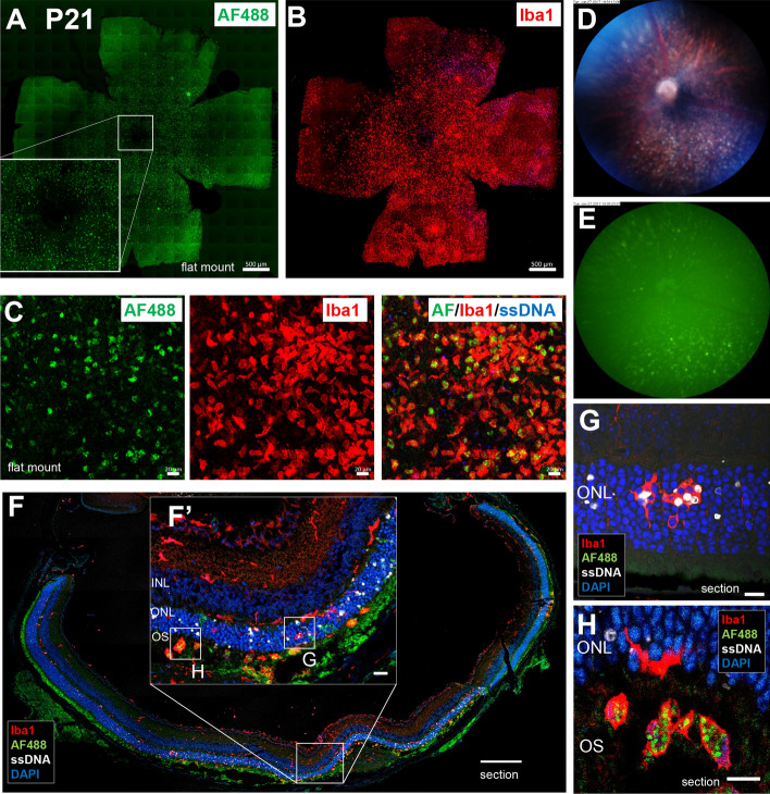
Figure 7.
Histopathological findings on diffuse fundus autofluorescent spots in the retina of rd10 at P21. (A) Accumulation of FAF spots was observed in the ONL around the central retina of rd10 at P21. Scale bar, 500 μm. (B) Accumulation of Iba1+ microglia was observed in the ONL layer around the centeral retina of the same mouse. Scale bar, 500 μm. (C) Flat-mounted retina of rd10 at P21 showing autofluorescent spots were labeled with Iba1 and ssDNA. The autofluorescent spots coincided with Iba1+ microglia but not with ssDNA+ apoptotic cells. Scale bars, 20 μm. (D) In the color fundus photograph, white granules accumulated under the retina were seen on P21. (E) In vivo FAF imaging of the same rd10 mouse. The localization of the fluorescent spots coincided with white granules in the subretina. (F) Retinal section of rd10 at P21 labeled with Iba1, ssDNA and DAPI. More apoptotic photoreceptors were observed in the ONL around the central retina. Scale bar, 200 μm. (F′) In retinal section near the center, Iba1+ microglia accumulated in the OS. Scale bar, 20 μm. (G) In the magnified image of the ONL, microglia that infiltrated into the ONL phagocytosed ssDNA positive and negative photoreceptors. Scale bar, 10 μm. (H) In the magnified image of the subretina, microglia in the OS stored fluorescent granules in their body. Scale bar, 10 μm.

