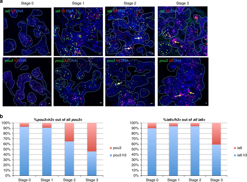
Fig. 2. Whole body regeneration is associated with proliferation of blood-borne integrin-alpha-6+ cells.
a Fluorescent in situ hybridization (FISH) showing expression of integrin-alpha-6 (ia6, green) and histone 3 (h3, red) during stages 0, 1, 2, and 3 of WBR. White arrows indicate ia6+ cells beginning to cluster during stage 2. As clusters increase in size, they begin to lose ia6 expression. As cell clusters differentiate to form the blastula-like structure, ia6 expression is not detected (yellow arrows). DNA was stained with Hoechst (blue) in all panels. Blue dashed lines outline blood vessel boundaries. Images are representatives of four independent experiments. Scale bars 20 μm. b Single-positive (ia6, pou3, or h3) and double-positive (ia6/h3 or pou3/h3) cells were counted using the cell counter feature in FIJI, and for each stage, four images from four independent samples were counted. Graph shows percentages of ia6/h3 double-positive or pou3/h3 double-positive cells among all ia6+ or pou3+ cells. Source data are provided as a Source Data file.

