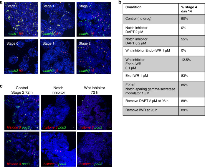
Fig. 6. Inhibition of Notch or Wnt signaling prevents WBR and blocks proliferation of pou3+ cells.
a Top panel: FISH for notch1 (green) and h3 (red) during steady state (stage 0) and during early stages of WBR. Bottom panel: FISH for notch2 (green) and h3 (red) during steady state (stage 0) and during early stages of WBR. Images are representatives of four independent experiments. Scale bar 20 μm. b Table showing the percentages of vessel fragments reaching stage 4 at day 14 post injury during treatment with different doses of inhibitors of canonical wnt signaling (Endo-IWR), Notch-signaling (DAPT) or control drugs Exo-IWR (1 μM) and the Notch-sparing gamma-secretase modulator E2012 (1 μM). The number of colonies that reached stage 4 was counted at 14 days post injury. Percentages represent the number of fragments reaching stage 4, with n = 14–18 fragments for each condition. Controls (subclones of the same genotype) received vehicle only. c FISH for pou3 (green) and h3 (red) showing a reduction in proliferating pou3+ cells 72 h after surgery when treated with an inhibitor of either Notch or Wnt signaling. Images are representatives of four independent experiments. Scale bars 20 μm.

