. 2020 Jul 6;142(8):081012. doi: 10.1115/1.4046974

Fig. 1.

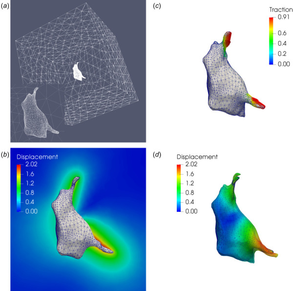
Microglial cell: (a) finite element mesh on the cell–gel interface and the surface of the truncating domain; (b) simulated tractions (in kPa) applied to the cell surface; (c) displacement field (in μm) on a 2D cross section of the hydrogel matrix; (d) displacement field (in μm) on the cell surface, where the solid and transparent surfaces represent the cell geometry in the stressed and relaxed configurations, respectively

Microglial cell: (a) finite element mesh on the cell–gel interface and the surface of the truncating domain; (b) simulated tractions (in kPa) applied to the cell surface; (c) displacement field (in μm) on a 2D cross section of the hydrogel matrix; (d) displacement field (in μm) on the cell surface, where the solid and transparent surfaces represent the cell geometry in the stressed and relaxed configurations, respectively