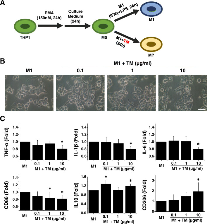
Fig. 1.
Thrombomodulin (TM) enhances M2 macrophage polarization in the presence of inflammatory cytokines. a A schematic diagram of the THP-1 induction protocol demonstrating the process of macrophage differentiation. b Representative light microscopy images revealed that TM-treated M1 macrophages exhibited no apparent change in morphology compared with M1 macrophages. Scale bar, 10 μm. c The expression levels of M1 and M2 markers were tested by using quantitative RT-PCR. The quantitative RT-PCR data demonstrated that the addition of TM to the M1 induction medium disrupted M1 polarization and enhanced polarization toward the M2 phenotype. n = 5. Mean ± SD. *p < 0.05 compared with M1

