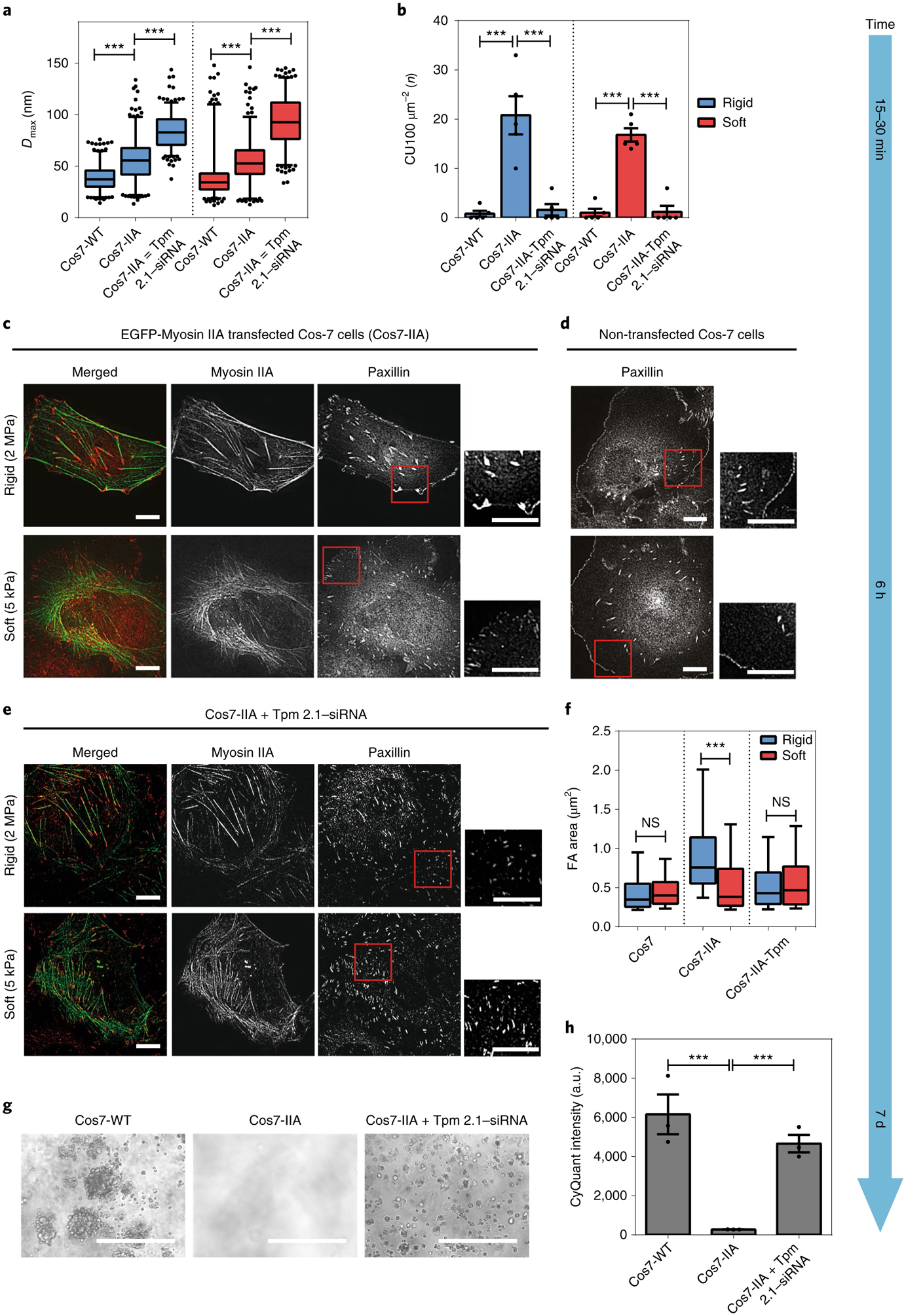
Fig. 3 |. Myosin IIA in Cos7 cells enables normal growth, but Tpm 2.1 silencing restores transformation.

a, Box-and-whisker plots of pillar Dmax for Cos7-WT, Cos7-IIA and Tpm-siRNA-transfected Cos7-IIA cells on rigid (blue) and soft (red) pillars. Silencing of Tpm 2.1 in Cos7-IIA cells increased the average force level on both types of pillar. b, Bar graphs of average CU density per 10 min for Cos7-WT, Cos7-IIA and Tpm 2.1–siRNA-transfected Cos7-IIA cells on rigid (blue) and soft (red) pillars (n = 5 in each case; error bars are s.e.m.; ***P < 0.001). c–e, Paxillin images of EGFP-myosin IIA-transfected Cos7 cells (Cos7-IIA) (c), Cos7 cells (d) and Tpm 2.1-depleted Cos7-IIA cells (e) fixed 6 h following seeding on rigid (2 MPa) or soft (5 kPa) fibronectin-coated PDMS surfaces (scale bars, 10 μm). f, Box-and-whisker plots of single focal adhesion area (FA) of Cos7, Cos7-IIA and Tpm 2.1-silenced Cos7-IIA cells on rigid (blue) and soft (red) PDMS surfaces (>150 adhesions were analysed in each case; ***P < 0.001). g,h, Soft agar assay indicating the growth of Cos7 and Tpm 2.1-knockdown Cos7 IIA cells, but not Cos7-IIA cells, after culture for 7 d (scale bars, 400 μm; n = 3 in each case; error bars are s.e.m.; ***P < 0.001; a.u., arbitrary units).
