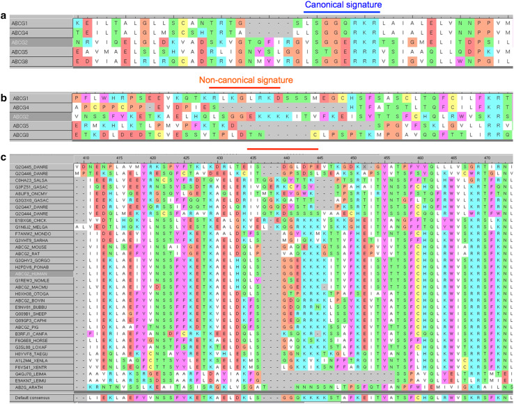
Fig. 1.
Sequence alignment of human ABCG sub-family members, emphasizing the regions containing the canonical signature/C-motif (marked with a blue line) (a) or a non-canonical C2-sequence that is only present in ABCG2 (marked with red line) (b). Alignment of the linker region containing the non-canonical C2-sequence of human ABCG2 with homologous regions of ABCG2 homologs from various species (c)

