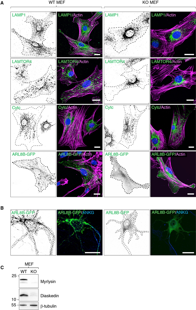
Figure 4. Perinuclear Clustering of Lysosomes and Dissociation of ARL8 in Cells from Myrlysin-KO Mice.

(A) MEFs from WT and KO E15 embryos were immunostained for endogenous LAMP1 and LAMTOR4 (lysosomes) or cytochrome c (Cytc) (mitochondria). Alternatively, MEFs were transiently transfected with a plasmid encoding ARL8B-GFP and immunostained for GFP. Cell edges were outlined by staining of actin with fluorescent phalloidin and indicated by the dashed lines. Nuclei were stained with DAPI. Scale bars: 20 μm.
(B) Hippocampal neurons from WT and KO E18 embryos were transfected with a plasmid encoding ARL8B-GFP at DIV 4 and immunostained for ANKG and GFP 1 day later. Cell edges are outlined by dashed lines. Scale bars: 20 μm.
(C) SDS-PAGE and immunoblot analysis of WT and KO MEFs using antibodies to myrlysin, diaskedin, and β-tubulin (loading control). The positions of molecular mass markers (kDa) are indicated on the left.
In (A) and (B), single-color images are represented in inverted grayscale.
