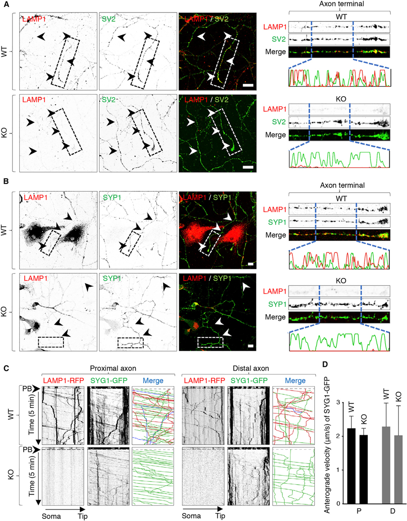
Figure 5. Distinct Effect of Myrlysin KO on the Localization and Movement of Lysosomes and SVPs in the Axon.

(A and B) DIV5 cultures of hippocampal neurons from WT and myrlysin-KO E18 embryos were immunostained for endogenous LAMP1 together with SV2 (A) or SYP1 (B). Images on the left show single-channel images in inverted grayscale and merged images in color. Arrowheads indicate the axon. Scale bars: 10 μm. Images on the right show straightened 50 μm segments of the distal axon taken from images on the left. Line intensity scans from 15 μm segments are also shown for LAMP1 and SV proteins.
(C) Straightened 50 μm segments of the proximal and distal axon from WT and myrlysin-KO E18 hippocampal neurons co-expressing LAMP1-RFP and SYG1-GFP that were analyzed at DIV5 by live-cell imaging and kymographs as described in the legend to Figure 1. Single-channel images are shown in inverted grayscale. In the merge panel, red (RFP) and green (GFP) lines represent moving vesicles having a single marker, and blue lines represent vesicles having both markers.
(D) Quantification of the anterograde velocity of SYG1-GFP-positive particles in the proximal (P) or distal (D) axon of WT and KO neurons. Values are mean ± SD of >100 vesicles in six neurons per condition and are not statistically different.
See also Figure S5.
