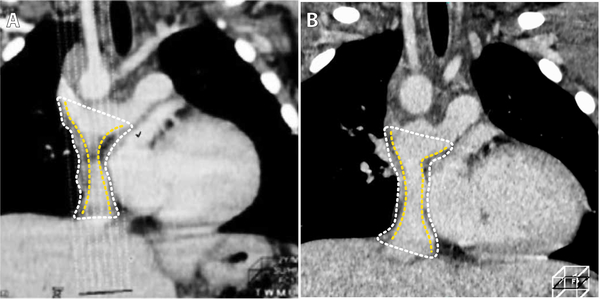
Fig. 7. Potential clinical case of reversible TEVG stenosis.
(A) CT images of the TEVG (outer wall outlined with white dots and lumen outlined with yellow dots) from patient 13 from the Japanese trial demonstrating wall thickening and luminal narrowing that presented during the first year after implantation and (B) resolved over the course of several months after treatment with oral anticoagulation (15).

