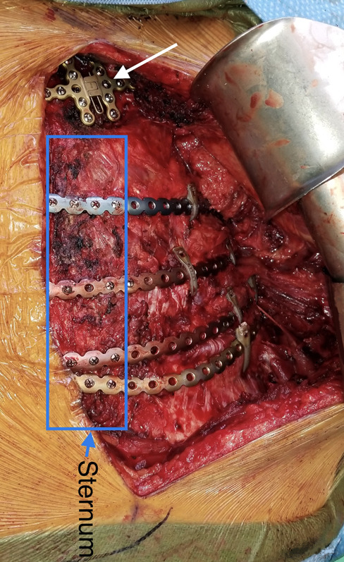
Fig. 11.

Intraoperative photograph showing multiple left-sided fractures of rib cartilage (ribs 3 to 6) stabilized by long threaded plates and cerclage fixation. Empty plate holes are seen above the fractured cartilage. The left pectoralis major muscle is retracted laterally. The plates are attached by screws medially to the sternum and laterally to the osseous part of the rib, with no screws through the cartilage. The manubrium fracture is stabilized by an H-shaped sternal plate (white arrow).
